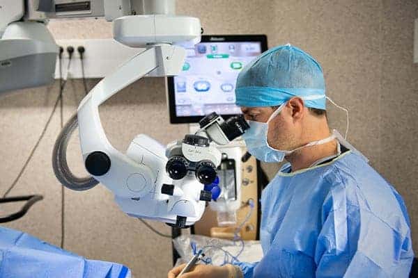
LASIK surgery in Paris
LASIK (Laser-Assisted In Situ Keratomileusis), performed entirely with a laser, is the most commonly used technique in refractive surgery, accounting for about two-thirds of all procedures in this field. The medical community has significant experience with this reference method, which enables the correction of myopia, astigmatism, hyperopia, and presbyopia.
Learn more about other refractive eye surgeries
Presbylasik
PRK
Trans PRK
Smile
What is LASIK surgery?
However, this is not always the case:
- In myopic (nearsighted) patients, the image forms in front of the retinal plane, resulting in impaired distance vision.
- Conversely, in hyperopic (farsighted) patients, images are formed behind the retina.
- Astigmatism occurs when different parts of the same image are not focused on the same plane, usually due to an irregular curvature of the cornea or lens.
- Presbyopia, which appears with age, results from a decrease in the lens’s ability to change shape for accommodation, making it difficult to see nearby objects.
LASIK is a surgical method that uses a laser beam to modify the curvature of the cornea, thereby changing the eye’s refractive power. This technique can correct the aforementioned vision disorders.
Find all Dr Rambaud’s videos on his Youtube channel.
@DocteurCamilleRambaud
LASIK for myopia, astigmatism, and hyperopia: indications
The preoperative assessment is essential, particularly to ensure that the patient has no contraindications to the treatment. Corneal thickness is a key factor, as the procedure involves working within the deeper layers of the stroma, located beneath the corneal epithelium and above the endothelium (the deepest layer).
Patients with excessively thin corneas should be excluded from the protocol and directed toward an alternative method, such as PRK, a surface laser technique.
If the patient is eligible, LASIK can treat mild to severe myopia (up to -8 diopters), hyperopia up to +5 diopters, and astigmatism up to 4 diopters.
A variant of the technique, PresbyLASIK, allows for the treatment of presbyopia.
LASIK: procedure
LASIK refractive surgery does not require hospitalization. The patient can return home the same day. Anesthesia is administered using anesthetic eye drops a few minutes before the procedure. In some cases, an anxiolytic may be given for additional comfort.
At the start of the procedure, a device is used to keep the eye open.
The first step is to cut a small flap at the surface of the cornea, which takes about twenty seconds using a femtosecond laser.
This “stromal flap” is then lifted to provide access to the stroma.
In the deeper layers, an excimer laser is used to make the necessary modifications to the cornea. This may involve flattening the center to treat myopia or thinning the peripheral cornea to make the central part bulge for hyperopia. The procedure is computer-assisted, with all parameters entered in advance, and an eye tracker ensures precision even if the eye moves.
At the end of the procedure, the stromal flap is repositioned. A bandage contact lens is then applied.
The entire operation takes no more than twenty minutes, even when both eyes are treated.
LASIK surgery: postoperative care
Postoperative instructions
Postoperative pain is moderate and more akin to a foreign body sensation in the eye, lasting only a few hours at most. Standard painkillers can be used if necessary. The prescribed treatment consists of antibiotic and anti-inflammatory eye drops for 15 days.
The procedure does not justify official sick leave, but it is advisable to plan for a 24-hour rest period.
The patient should avoid rubbing their eyes during the first week. Protective shields should be worn at night and glasses during the day throughout this period.
At least 7 days should pass before resuming physical activity, or longer if there is a risk of ocular trauma. Additionally, swimming should be avoided for the first 3 weeks.
Eyelids and eyelashes can be re-applied with makeup 10 days after the procedure, but eyeliner should not be used for at least 1 month.
LASIK: visual recovery and postoperative course
Postoperative complications are extremely rare.
However, temporary dry eye is quite common and can be managed with artificial tears. Sometimes, patients may experience increased light sensitivity, which is also temporary.
Vision returns very quickly after LASIK, but it may be blurry for a few hours. Significant improvement occurs after 12 hours, but the final result is only apparent once healing is complete, sometimes after several weeks. This timeframe depends on the degree of correction and the extent of the procedure.
LASIK LASER EYE SURGERY : PRICES IN PARIS
LASIK results
LASIK typically yields remarkable results, with significant improvement in the patient’s vision. However, glasses may still be needed in some cases, or a secondary procedure may be considered to refine the initial result.
Book an appointment with Dr. Rambaud
Frequently asked questions about LASIK
Why is a minimum corneal thickness required for LASIK?
In patients of Western origin, the average corneal thickness is 550 micrometers, including the epithelium (the most superficial layer), the stroma (intermediate layer), and the endothelium (deepest layer). The stromal flap created at the beginning of the procedure is about 120 micrometers thick (50 micrometers of epithelium, the rest stroma).
After the procedure, even though the stromal flap is repositioned, its thickness no longer contributes to the cornea’s mechanical stability. Stability is considered sufficient only with an effective thickness of 300 micrometers. Thus, the surgeon has 130 micrometers of stroma available for excimer laser photoablation (550 initial thickness – 120 stromal flap – 300 to preserve for stability).
Therefore, the higher the refractive error, the more stromal tissue must be removed. Some severe vision defects cannot be treated with LASIK because the required ablation would compromise safety.
What is keratoconus, and why is it a contraindication for LASIK?
Keratoconus weakens the cornea and is thus a strict contraindication for LASIK. Detecting it during the preoperative assessment is crucial.
Keratoconus is a rare, degenerative condition of still poorly understood origin, affecting 4 to 6 per 100,000 individuals according to studies. It is characterized by thinning of the corneal layers, which gradually take on an irregular cone shape.
It is most often detected during adolescence, especially when myopia or astigmatism progresses abnormally rapidly. Symptoms include visual fatigue, headaches, blurred vision, multiple or distorted images, poor distance vision, streaks, and increased light sensitivity. Complications can be serious, including corneal ulcers or perforations, stromal edema, and scarring.
In early stages, keratoconus can be managed medically with glasses or, more commonly, specialized contact lenses. Surgical treatment becomes necessary in more advanced cases. Options include corneal cross-linking (strengthening the cornea by removing the epithelium, applying vitamin B2, and irradiating with ultraviolet light) or the insertion of intracorneal rings. In all cases, the goal is to delay corneal transplantation as long as possible.
Have a question? Ask Dr. Rambaud
This page was written by Dr. Camille Rambaud, an ophthalmologist based in Paris and a specialist in refractive surgery.


0 Comments